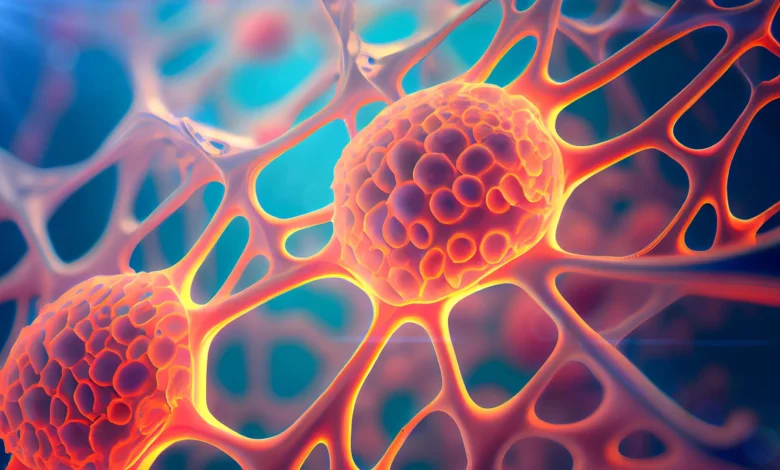
The human body possesses a remarkable, yet until recently mysterious, capacity to recover from catastrophic physical damage. Tissues spanning the skin, the gastrointestinal tract, and the protective epithelial layers encasing vital organs can routinely rebuild themselves after enduring severe trauma. For approximately fifty years, biologists have acknowledged this restorative phenomenon—formally termed compensatory proliferation—yet the precise intracellular triggers governing such dramatic cellular regrowth remained elusive. Now, groundbreaking research originating from the Weizmann Institute of Science in Israel has unraveled a crucial piece of this biological puzzle, revealing a paradoxical cellular mechanism where enzymes traditionally responsible for destroying cells instead orchestrate their survival and subsequent tissue repair.
Published in the peer-reviewed journal Nature Communications, the study sheds unprecedented light on the dual nature of caspases, a family of protease enzymes historically categorized as the executioners of programmed cell death, or apoptosis. According to the international research team, these enzymes can actively confer death resistance upon certain cells facing lethal stress. While this evolutionary safeguard enables severely damaged tissues to regenerate successfully, it simultaneously exposes a darker physiological vulnerability. Malignant cells may hijack this exact molecular survival pathway, potentially explaining why certain aggressive tumors endure aggressive therapies and return in treatment-resistant forms.
A Half-Century Mystery: Tracing the Roots of Compensatory Proliferation
The scientific journey toward understanding compensatory proliferation began in the 1970s. During this era, pioneer geneticists exposed fruit fly (Drosophila melanogaster) larvae to high dosages of ionizing radiation. Despite sustaining massive, widespread damage to their epithelial tissues, the irradiated larvae demonstrated a startling capacity to regenerate fully functional structures, such as adult wings. Over the subsequent decades, analogous regenerative responses were documented across a wide phylogenetic spectrum, eventually extending to mammalian systems, including humans.
Despite widespread observation of the phenomenon, the underlying mechanics baffled researchers. How could tissue heavily decimated by trauma manage not only to clear away dead debris but also to stimulate surviving cells to proliferate at precisely the correct rate to fill the void? Traditional biological dogmas viewed cell death and cell division as mutually exclusive pathways: a dying cell was simply discarded, and neighboring healthy cells passively divided to replace the loss.
The paradigm began to shift gradually over the past twenty years as global research groups—including the laboratory of Professor Eli Arama within the Department of Molecular Genetics at the Weizmann Institute of Science—began identifying nonlethal functions for apoptotic caspases. Arama, a leading figure in the investigation of these alternative caspase roles, hypothesized that these enzymes might act as the missing link driving compensatory proliferation. To test this hypothesis, a research team led by Dr. Tslil Braun in Arama’s laboratory set out to recreate the classic 1970s irradiation experiments using contemporary genetic tracing tools capable of resolving cellular dynamics at an unprecedented level of detail.
Decoding the Cellular Rescuers: DARE and NARE Populations
To observe how cells respond to lethal stress in real time, the Weizmann researchers engineered a sophisticated experimental model using Drosophila larvae. By deploying a specialized delayed sensor system, the team was able to track cells that activated their initiator caspases—the molecular trigger for apoptosis—yet miraculously survived the radiation onslaught.
"We set out to identify cells that push the self-destruct button but survive anyway," explained Dr. Tslil Braun, detailing the methodological approach. "To do this, we used a delayed sensor that reported on cells in which the initiator caspase had been activated but that nevertheless survived the irradiation. This is how we discovered a population of cells we named DARE cells."
The acronym DARE stands for Death-Apoptozic-Resistant-Epithelial cells, reflecting their unique origin story. Rather than perishing as programmed, these cells halted their destruction midway, survived the toxic radiation, and subsequently underwent rapid proliferation. Within a 48-hour window, DARE cells actively repaired the damaged epithelial landscape, single-handedly replenishing nearly half of the lost tissue.
However, this discovery introduced a mathematical and biological riddle: if DARE cells accounted for roughly 50 percent of the regenerated tissue, where did the remaining half originate? Further investigation uncovered a second, distinct population of death-resistant cells that contributed to the recovery effort. Unlike their DARE counterparts, these cells had never initiated the apoptotic cascade. The researchers designated this second group NARE cells (Non-Apoptotic-Resistant-Epithelial cells).
"We identified another population of death-resistant cells, but unlike DARE cells, they showed no activation of the initiator caspase. We called them NARE cells," Braun noted. Despite lacking the dramatic rescue history of DARE cells, NARE cells proved essential to the overarching recovery. The two populations operated interdependently; when researchers experimentally depleted the system of DARE cells, compensatory proliferation ceased entirely. Furthermore, dying neighboring cells emitted chemical distress signals that served as a catalyst, waking up and mobilizing the DARE cells to initiate their rescue mission.
Mechanistic Breakdown: Halting the Executioner
The research team then probed the precise molecular machinery responsible for allowing DARE cells to evade an apparent death sentence. Biochemical analysis revealed that the apoptotic pathway in DARE cells begins normally. The initiator caspase switches on, responding to the radiation-induced trauma. However, at a critical juncture, the signaling cascade stalls abruptly before effector (executioner) caspases can be recruited to dismantle the cell’s internal structural proteins.
According to Professor Arama, this arrest is mediated by specialized regulatory proteins. "We observed that although the initiator caspase is activated in these cells, the cellular death process stops there and does not progress to the next stage," Arama stated. "We suspected that a protein known as a molecular motor was responsible for this—it can tether the initiator caspase to the cell membrane, preventing it from activating the executioner caspases."
To confirm this hypothesis, the scientists silenced the gene encoding the molecular motor protein. When this tethering mechanism was disrupted, DARE cells could no longer halt their apoptotic cascade; they proceeded to die, and overall tissue regeneration was severely impaired.
Crucially, the implications of this molecular brake extend far beyond basic fruit fly physiology. Overactivation of identical motor proteins has previously been documented in human cancerous tumors. This structural parallel strongly suggests that cancer cells exploit this exact evolutionary safeguard, utilizing molecular tethers to evade radiation-induced apoptosis and survive aggressive medical interventions.
The Biological Legacy of Survival: Inherited Resistance
One of the most clinically alarming findings of the Weizmann study concerns the long-term biological consequences for cells that endure and survive primary trauma. Clinical oncologists have long observed that human tumors recurring after radiation therapy frequently exhibit heightened aggressiveness and profound resistance to subsequent rounds of treatment. To determine whether this acquired resilience is heritable, the Weizmann team evaluated the descendants of DARE cells.
"We wanted to understand whether resistance to death is inherited by the descendants of death-resistant cells that survived the initial irradiation," Professor Arama explained.
When the regenerated epithelial tissue was subjected to a second dose of ionizing radiation, the physiological response was strikingly different from the first exposure. The number of cells dying during the immediate post-irradiation window was reduced by half compared to the initial trauma, and the vast majority of those dying cells belonged to the vulnerable NARE population. Conversely, the descendants of the original DARE cells exhibited exceptional resilience—demonstrating a survival capacity seven times greater than standard, unexposed cells.
This inherited trait provides a compelling mechanistic explanation for clinical observations in oncology. When patients undergo radiotherapy, a fraction of the tumor cells may activate DARE-like survival pathways. While the immediate tumor burden shrinks, the surviving cells and their progeny inherit a profound resistance to cell death, rendering subsequent therapeutic interventions significantly less effective.
Maintaining Equilibrium: The Negative-Feedback Loop
While rapid and robust cellular proliferation is vital for closing open wounds and restoring organ integrity, unchecked cellular growth carries existential risks for an organism. Unregulated division is the foundational hallmark of cancer. Consequently, biological repair systems must incorporate rigorous stop-signals to prevent reparative processes from spiraling into runaway tumors.
In the final phase of their study, the researchers uncovered a sophisticated communication network operating between DARE and NARE cells that maintains this delicate homeostasis. The two populations do not merely coexist; they engage in a continuous biochemical dialogue characterized by reciprocal regulation.
"DARE cells promote the growth of nearby NARE cells, apparently by secreting growth signals," Arama observed. "In turn, NARE cells secrete signals that inhibit the growth of DARE cells. In fact, we’ve discovered a negative-feedback loop between the two cell populations that prevents overgrowth."
This intricate cross-talk ensures that once the epithelial gap is bridged and tissue architecture is restored, the proliferative burst is safely quenched, averting the development of hyperplastic lesions or accidental tumorigenesis.
Collaborative Effort and Institutional Leadership
This comprehensive investigation was conducted through an international, multidisciplinary collaboration. Alongside Professor Arama and Dr. Tslil Braun, the research team included Naama Afgin, Dr. Lena Sapozhnikov, and Dr. Keren Yacobi-Sharon from the Weizmann Institute’s Department of Molecular Genetics. Operational support and advanced technical infrastructure were provided by Dr. Ehud Sivan from Weizmann’s Life Sciences Core Facilities Department.
The study also benefited from vital international partnerships, featuring contributions from Prof. Andreas Bergmann of the UMass Chan Medical School in Worcester, Massachusetts, and Prof. Luis Alberto Baena-Lopez from the Severo Ochoa Molecular Biology Center in Spain. Professor Eli Arama currently serves as the incumbent of the Harry Kay Professorial Chair of Cancer Research and directs the Crown Human Genome Center at the Weizmann Institute.
Broader Implications for Regenerative Medicine and Oncology
While the current experiments were performed using Drosophila models, historical precedent strongly supports the translatability of these findings to human biology. Fundamental signaling pathways governing apoptosis and cellular proliferation are deeply conserved across evolutionary history, and discoveries in invertebrate models have frequently paved the way for breakthroughs in human medicine.
The dual nature of the DARE-NARE survival system presents both a formidable challenge and an inspiring opportunity for modern biomedical science. On one hand, understanding how caspases can be repurposed for cell survival rather than execution opens up novel therapeutic avenues in regenerative medicine. By safely pharmacologically mimicking or stimulating DARE-like mechanisms, clinicians might one day accelerate the healing of chronic wounds, severe burns, or degenerative tissue damage in human patients.
Conversely, the same molecular insights offer a rational framework to combat oncology’s most persistent frustration: cancer recurrence. If pharmaceutical researchers can develop targeted inhibitors against the molecular motors or tethering proteins that allow cancer cells to mimic DARE survival pathways, they may strip tumors of their acquired death resistance. This approach could sensitize treatment-refractory cancers to conventional radiotherapy and chemotherapy, ensuring that cancer cells cannot turn their self-destruct mechanisms off and return stronger than before.
Ultimately, the Weizmann Institute study bridges two traditionally disparate fields of biomedicine—regenerative repair and cancer biology. By illuminating the fine line that separates healthy tissue healing from malignant evasion, these findings provide a foundational roadmap for developing advanced therapies that promote vital tissue recovery while denying deadly diseases the tools they need to survive.