New preclinical research from The University of Texas MD Anderson Cancer Center has uncovered a sophisticated biological partnership between short-term fasting and a specific gut microbe, demonstrating how dietary changes can prime the body’s natural tissue repair mechanisms. Published in the Proceedings of the National Academy of Sciences, the study reveals that a 24-hour fast conditions intestinal cells to recover more rapidly from radiation damage, but this protective mechanism is entirely dependent on the presence of a beneficial bacterium known as Akkermansia muciniphila.
The findings illuminate a complex network of interactions bridging nutrition, the human microbiome, and cellular epigenetics. While the research remains in the preclinical phase, it opens new avenues for mitigating the collateral damage of cancer treatments, potentially improving the safety and efficacy of radiotherapy for abdominal tumors. By detailing how dietary interventions and microbially produced metabolites synergize to alter gene expression in intestinal stem cells, the research team has laid the groundwork for future clinical strategies designed to protect vulnerable healthy tissues without necessarily requiring patients to undergo rigorous fasting regimens.
Understanding the Vulnerability of the Gastrointestinal Tract in Cancer Therapy
Radiation therapy remains a cornerstone of modern oncology, deployed extensively to target and eradicate malignancies in the abdominal and pelvic cavities, including pancreatic, colorectal, and gynecologic cancers. Despite continuous technological advancements in precision targeting, high-energy ionizing radiation inevitably intersects with surrounding healthy tissue. The small intestine is notoriously vulnerable to these therapeutic beams. Because the lining of the small intestine features a rapid cellular turnover rate to maintain its barrier function, its delicate epithelial cells are hypersensitive to radiation-induced DNA damage.
When radiation destroys this protective mucosal barrier, patients frequently experience a cascade of acute and debilitating side effects, ranging from severe nausea, cramping, and debilitating diarrhea to systemic infections. In extreme cases, radiation enteritis can lead to life-threatening complications such as bowel perforation, sepsis, and chronic malabsorption. These acute toxicities impose strict clinical limitations on the cumulative radiation dose oncologists can safely deliver to a tumor site. Consequently, the fear of severe gastrointestinal toxicity can occasionally compromise the curative potential of cancer treatment.
For decades, clinical radiobiology has sought methods to selectively shield healthy tissue while maximizing the cytotoxic payload delivered to the tumor. While various pharmacological radioprotectors have been investigated, few have achieved widespread clinical adoption due to toxicity concerns or the risk of simultaneously protecting the cancer cells. This background sets the stage for investigations into endogenous physiological safeguards, such as caloric restriction, which may exploit the body’s natural resilience pathways.
Tracing the Chronology: From Laboratory Curiosity to Mechanistic Discovery
The trajectory of this research stems from earlier investigations conducted within the Piwnica-Worms Laboratory at MD Anderson Cancer Center. In antecedent preclinical studies, researchers observed a striking phenomenon: laboratory models subjected to a brief, 24-hour fast prior to receiving high-dose abdominal radiation exhibited a significantly enhanced capacity for intestinal recovery compared to non-fasting counterparts. The fasted subjects suffered less mucosal destruction and regenerated their intestinal lining at an accelerated rate.
However, observing the protective effect of fasting and understanding its exact molecular etiology are two entirely different scientific hurdles. While general metabolic shifts—such as reduced systemic glucose, alterations in growth factor signaling, and the induction of cellular autophagy—were known to accompany short-term caloric restriction, these factors alone could not fully account for the robust, targeted tissue regeneration observed in the gut.
Recognizing that the gastrointestinal tract houses trillions of microorganisms that dynamically respond to dietary shifts, the research team shifted their focus toward the gut microbiome. Co-led by Dr. Helen Piwnica-Worms, professor of Experimental Radiation Oncology, and Dr. Kunal Rai, professor of Genomic Medicine, the team hypothesized that specific microbial populations might act as mandatory mediators of the fasting-induced radio-protective effect. Through rigorous microbial profiling and targeted depletion experiments over the subsequent research cycle, the investigators isolated the critical microbial player: Akkermansia muciniphila, commonly abbreviated as AKK.
The Biological Cascade: How Fasting and Microbes Instruct the Gut
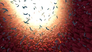
The newly published study maps out the precise sequence of events that unfolds when a 24-hour fast is combined with the metabolic activity of Akkermansia muciniphila. The process begins in the lumen of the small intestine, where a temporary cessation of food intake selectively enriches the population of AKK. This mucin-degrading bacterium thrives under specific nutrient-deprived conditions and produces metabolic byproducts, most notably the short-chain fatty acid propionate.
Propionate does not merely act as an energy source; it functions as a potent signaling molecule that penetrates the epithelial cells lining the intestine. Inside these cells, propionate acts in concert with broader metabolic alterations triggered by fasting to induce profound epigenetic modifications. Specifically, these combined signals alter histones—the protein spools around which genomic DNA is tightly wound.
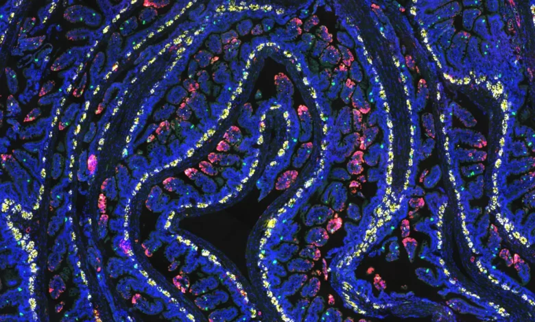
By adding small chemical tags to these histone proteins, the cell alters its chromatin architecture without changing the underlying genetic code. Certain sections of DNA that dictate tissue regeneration and stem cell activation become structurally uncoiled and accessible. Consequently, a specific population of intestinal cells is pre-programmed with an emergency preparedness plan.
"Fasting helps prepare intestinal cells to respond more quickly and effectively after injury, almost like training the cells with an emergency preparedness plan," explained Dr. Helen Piwnica-Worms. "This study helps explain how that plan is organized and identifies a key bacterium involved in coordinating the response."
When the intestines are subsequently exposed to ionizing radiation, these epigenetically primed cells do not succumb to apoptosis as easily as unprimed cells. Instead, they rapidly proliferate and expand, orchestrating a swift and efficient rebuilding of the damaged mucosal barrier.
The Indispensable Role of Akkermansia Muciniphila
To definitively prove that Akkermansia muciniphila was a non-negotiable component of this biological chain reaction rather than a mere bystander, the researchers performed targeted depletion assays. By selectively eliminating AKK from the microbiome of the experimental models, the protective benefits of the 24-hour fast were completely abrogated. Under conditions lacking AKK, fasted subjects suffered the same degree of severe radiation-induced intestinal damage as non-fasted subjects.
Crucially, when the researchers exogenously restored Akkermansia muciniphila to the microbial ecosystem of the depleted subjects and paired it with the 24-hour fast, the robust tissue recovery phenotype was fully rescued. This control experiment provided unequivocal evidence that neither fasting alone nor the presence of the bacterium alone is sufficient to maximize tissue resilience. The phenomenon requires a synergistic collaboration between host caloric restriction and microbial metabolism.
This discovery reframes how scientists view the gut microbiome during therapeutic interventions. Rather than viewing bacteria as passive inhabitants that merely assist in digestion, the findings position specific microbes as active co-regulators of host stress responses and cellular regeneration.
Implications for Oncology and Future Clinical Horizons
While the implications of this discovery are profound, the research team and independent oncologists emphasize that the findings are currently limited to preclinical models. Translating these insights into human clinical practice will require rigorous clinical trials to confirm whether human intestinal tissue responds to fasting and AKK modulation in an identical manner.
Nevertheless, the potential clinical applications are expansive. Abdominal radiation therapy affects hundreds of thousands of cancer patients annually. If these mechanistic pathways can be safely and reliably replicated in humans, the medical community could witness a paradigm shift in how radiation toxicities are managed.
Furthermore, the practical challenges of enforcing a strict 24-hour fast on cancer patients—many of whom are already experiencing systemic fatigue, weight loss, and cachexia—cannot be overstated. Recognizing this hurdle, the research team is already looking toward translational alternatives that bypass the need for patient food restriction altogether.
"Fasting is not always practical for cancer patients, and this work supports several other potential ways to enhance recovery after treatment," noted Dr. Kunal Rai. "Whether through dietary interventions, targeted microbes or their metabolites, the goal is to help repair healthy tissue more effectively while patients receive the cancer therapies they need."
Future therapeutic explorations may involve administering targeted prebiotics to naturally stimulate indigenous AKK populations, utilizing live biotherapeutic products containing formulated strains of Akkermansia muciniphila, or directly delivering microbial metabolites like propionate to patients undergoing radiotherapy. Additionally, researchers are eager to investigate whether this same fasting-microbiome axis can be harnessed to protect other rapidly dividing healthy tissues vulnerable to cancer therapy, such as bone marrow progenitor cells.
Funding and Institutional Support
This complex interdisciplinary study was supported by major institutional grants, reflecting its high priority within the cancer research landscape. Primary financial backing was provided by the National Cancer Institute (NCI), a division of the National Institutes of Health (NIH), alongside critical support from the Cancer Prevention and Research Institute of Texas (CPRIT).
As MD Anderson Cancer Center researchers move toward the next phases of investigation, the scientific community awaits translational studies that will test whether harnessing the power of a single gut microbe can ultimately make cancer therapy safer and more tolerable for patients worldwide.