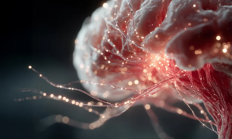
The architecture of the human brain has long been studied through the isolated lens of gray matter degradation, viewing cognitive decline primarily as a casualty of neuronal cell loss. However, groundbreaking new research led by scientists at the Mark and Mary Stevens Neuroimaging and Informatics Institute (Stevens INI) at the Keck School of Medicine of the University of Southern California (USC) suggests that the brain’s local communication pathways play a vital, previously underappreciated role in preserving cognitive function in older adults. Their findings indicate that the structural integrity of the brain’s microscopic local wiring—specifically superficial white matter—can act as a biological buffer, potentially mitigating the cognitive fallout typically associated with the loss of gray matter.
Published in the peer-reviewed journal Alzheimer’s & Dementia: The Journal of the Alzheimer’s Association, the study evaluates brain imaging and cognitive assessments derived from a diverse, community-based cohort of 459 adults aged 60 and older. Crucially, the participants reside across various communities in India, marking this research as a pioneering effort to study superficial white matter within an underrepresented population from a low- and middle-income country. By looking beyond traditional Western demographic samples and expanding the methodological scope to include the brain’s hidden local networks, the USC-led team has opened new avenues for understanding cognitive resilience, brain aging, and the complex interplay between physical neural infrastructure and environmental life factors.
Decoding the Brain’s Local Communication Network
To understand the mechanics of the study, one must examine the distinct yet complementary roles played by the brain’s primary tissue types. Gray matter, which forms the outer cortex of the brain, is dense with nerve cell bodies responsible for processing information, computing decisions, and executing conscious thought. Directly beneath this cortical mantle lies a thin, intricate layer of short, curved nerve fibers known as superficial white matter.
If gray matter acts as the urban centers where intellectual processing takes place, superficial white matter functions as the network of local roads connecting adjacent neighborhoods. These short-range fibers facilitate rapid communication between neighboring regions of the cerebral cortex, allowing them to share data seamlessly. While long-range white matter tracts connect distant lobes of the brain—acting like interstate highways—superficial white matter governs localized, highly integrated regional processing.
"Gray matter and superficial white matter are physically close and may play different roles: gray matter processes information, while superficial white matter helps nearby brain regions communicate," explained Yingxu Liu, PhD, a postdoctoral scholar at the Stevens INI and first author of the study. "Our findings suggest that cognitive health depends not only on how much gray matter is preserved, but also on the condition of the wiring that connects it."
This interdependence implies that cognitive vitality is a systemic property rather than the product of any single isolated region. When the local wiring is robust, adjacent brain regions can route around minor inefficiencies or compensate for localized micro-structural damage. Conversely, when both the processing centers (gray matter) and the local roads (superficial white matter) degrade simultaneously, the system experiences catastrophic failures in communication, manifesting clinically as measurable cognitive decline.
Advanced Imaging Techniques Reveal Microscopic Wiring
Investigating superficial white matter has historically presented a significant challenge for neuroscientists. Because these fibers are short, curved, and densely packed immediately beneath the convoluted folds of the cerebral cortex, conventional brain scans often lack the resolution required to isolate them from adjacent tissues. To overcome this hurdle, the research team utilized an advanced modality of diffusion magnetic resonance imaging (MRI).
Diffusion MRI tracks the micro-scale diffusion of water molecules through biological tissues. By measuring how water molecules move along restricted pathways dictated by nerve cell boundaries, scientists can infer the micro-structural health of the tissue without invasive procedures. Specifically, the researchers focused on metrics quantifying neurite density and free-water fraction.
Neurites are the minute axonal and dendritic projections through which neurons transmit and receive electrical and chemical signals. A reduction in neurite density, coupled with an elevated volume of freely moving water in the extracellular space, serves as a reliable biomarker for tissue disruption, micro-structural degeneration, myelin breakdown, neuroinflammation, or cellular edema. By mapping these minute variations, the USC team gained an unprecedented look into the physical condition of the brain’s local communication network.
Parallel to these advanced scans, participants completed comprehensive cognitive evaluations. These assessments measured multiple functional domains, including language faculties, memory retention, executive functioning, and visuospatial orientation. Statistical modeling was subsequently applied to determine how variations in superficial white matter integrity correlated with performance across these cognitive domains.
The Lingering Power of Language and Frontotemporal Resilience
Among the various cognitive domains tested, language performance emerged as the most consistent and prominent correlate of superficial white matter health. Participants with higher structural integrity in their superficial white matter consistently scored higher on language-related tasks. Furthermore, the strongest associations were concentrated within the frontotemporal regions of the brain—cortical areas intimately involved in semantic word recognition, speech fluency, and verbal working memory.
While measurements of gray matter atrophy remained the single strongest overall predictor of general cognitive capability across the cohort, the health of the superficial white matter introduced a critical moderating variable. The data revealed that the clinical impact of gray matter loss is not uniform across all individuals; rather, its severity depends heavily on the condition of the local wiring.
When superficial white matter showed advanced deterioration or low structural integrity, the negative association between gray matter loss and impaired cognitive performance—particularly in language tasks—was pronounced and severe. However, when the superficial white matter remained healthy and intact, the detrimental link between gray matter atrophy and cognitive impairment was substantially attenuated.
This buffering effect offers a compelling biological explanation for a long-standing clinical puzzle: why two individuals experiencing identical degrees of cortical gray matter shrinkage can exhibit drastically different levels of cognitive sharpness in their daily lives.
"The findings point to superficial white matter as a possible source of resilience," noted Leon Aksman, PhD, assistant professor of research neurology at the Stevens INI and senior author of the study. "Two people with a similar degree of gray matter loss may not experience the same cognitive effects if the local connections surrounding that gray matter differ in health. Following participants over time will be essential to test whether preserving these connections can help maintain cognition."
Broadening Horizons: Inclusion of Underrepresented Populations
A defining characteristic of this research lies in its demographic foundation. The data analyzed by the USC team was drawn from the Harmonized Diagnostic Assessment of Dementia for the Longitudinal Aging Study in India, widely known as LASI-DAD. Historically, neuroimaging and cognitive aging studies have suffered from a severe Western-centric bias, predominantly recruiting urban, highly educated populations from high-income nations.
The LASI-DAD cohort drastically expands this horizon. More than half of the broader LASI-DAD participant pool exhibits low literacy rates, and approximately 60% reside in rural communities across India. By analyzing brain imaging from this diverse socioeconomic and geographic demographic, the researchers were able to investigate brain aging across a spectrum of life experiences that rarely feature in mainstream neuroscientific literature.
Intriguingly, the association between superficial white matter health and language ability was found to be even stronger among participants who were functionally illiterate or had received no formal education, as well as those living in rural environments.
The authors are careful to interpret these correlations cautiously. They emphasize that these sociological factors do not directly cause specific micro-structural changes in brain tissue in a simplistic manner. Instead, the results suggest that brain aging is molded by a cumulative, lifelong matrix of environmental exposures, educational opportunities, socioeconomic circumstances, systemic health conditions, and lifestyle factors. This reinforces the necessity of building neuroscientific models that reflect the true diversity of the global population.
Implications for Future Alzheimer’s and Dementia Research
Despite the significance of these cross-sectional findings, the study’s design carries inherent limitations that point the way forward for subsequent research. Because the participants were assessed at a single point in time, the study cannot definitively establish temporal precedence. Scientists cannot yet determine whether the deterioration of superficial white matter acts as an initial trigger that subsequently accelerates gray matter loss, whether the two processes unfold simultaneously, or whether micro-structural white matter breakdown occurs strictly as a downstream consequence of cortical atrophy.
To untangle these complex temporal dynamics, longitudinal studies tracking individuals over extended periods of time will be essential. Future phases of this research agenda will focus on monitoring how these structural brain changes evolve as participants age, while also integrating biological variables such as vascular health markers, systemic inflammation levels, and the accumulation of Alzheimer’s-related neuropathological proteins like amyloid-beta and tau.
Arthur W. Toga, PhD, director of the Stevens INI and Provost Professor at USC, underscored the broader implications of the work for the global scientific community. "A fuller understanding of brain aging requires research that reflects the world’s social, cultural, and geographic diversity," Toga stated. "By studying an underrepresented population and looking beyond gray matter alone, this work brings us closer to identifying the biological and social factors that may protect cognition across the lifespan."
The study’s collaborative authorship reflects a vast, multidisciplinary effort spanning international research institutions. In addition to Liu and Aksman, the author roster features Kirsten M. Lynch, Miguel Arce Rentería, Emma Nichols, Alden L. Gross, Lindsay C. Kobayashi, Neda Jahanshad, John P. John, Harshita V. Vishwakarma, Pranali Khobragade, Joyita Banerjee, Niranjan Khandelwal, Jyoti Dangwal, Sudhir Saxena, Nirod Medhi, Soumik Das, Prudhvinath Reddy, Pratyaksha Rana, Arjun Narula, Saravanan Kannan, Dinesh Patel, A. B. Dey, Sharmistha Dey, and Jinkook Lee.
Financial backing for the expansive research initiative was provided by several major branches of the United States National Institutes of Health, including the National Institute on Aging (grants R01AG080473, RF1AG087965, RF1AG088003, and R01AG087513), the National Institute of Mental Health (grant R01MH134004), the National Institute of Neurological Disorders and Stroke (grant RF1NS136995), and the Office of the Director of the National Institutes of Health (grant S10OD032285).
As the global population ages and the prevalence of neurodegenerative conditions continues to mount, shifting the medical research paradigm from mere disease tracking toward the identification of endogenous resilience factors represents a vital evolution. By illuminating the protective capacity of superficial white matter, this USC-led study paves the way for novel therapeutic and preventative interventions aimed not just at protecting brain cells, but at preserving the intricate, vital roadways that allow the human mind to communicate with itself.